

Mycelium of Mycena rubromarginata cultured in the lab. A mycelium is a network of fungal threads. It grows underground or in other places such as rotting tree trunks, twigs and fallen leaves. The fruiting bodies of fungi, such as mushrooms, can sprout from a mycelium. Mycelia are vital for the health of soil in many ecosystems. They break down organic material making it available for other organisms and sustaining continuous growth.

An Evolutionary Trap. Depicted is the balanced lethal system in Triturus salamanders, an example of a bizarre, evolutionary trap. The hereditary disease consistently kills half the reproductive output and likely originated dozens of millions of years ago in Triturus’ most recent common ancestor, who I like to call ‘patient zero’.

I used computed tomography (CT) images of the Visible Human Project®, specifically the Visible Human Female to reconstruct the full body of the female. The data representing the patient was isolated from the background and one in every 18 slices was depicted to reconstruct a visualisation of the full body. I added a marble base to create a more tangible effect. Our goal is to print each slice on glass and reconstruct the patient in life-size.
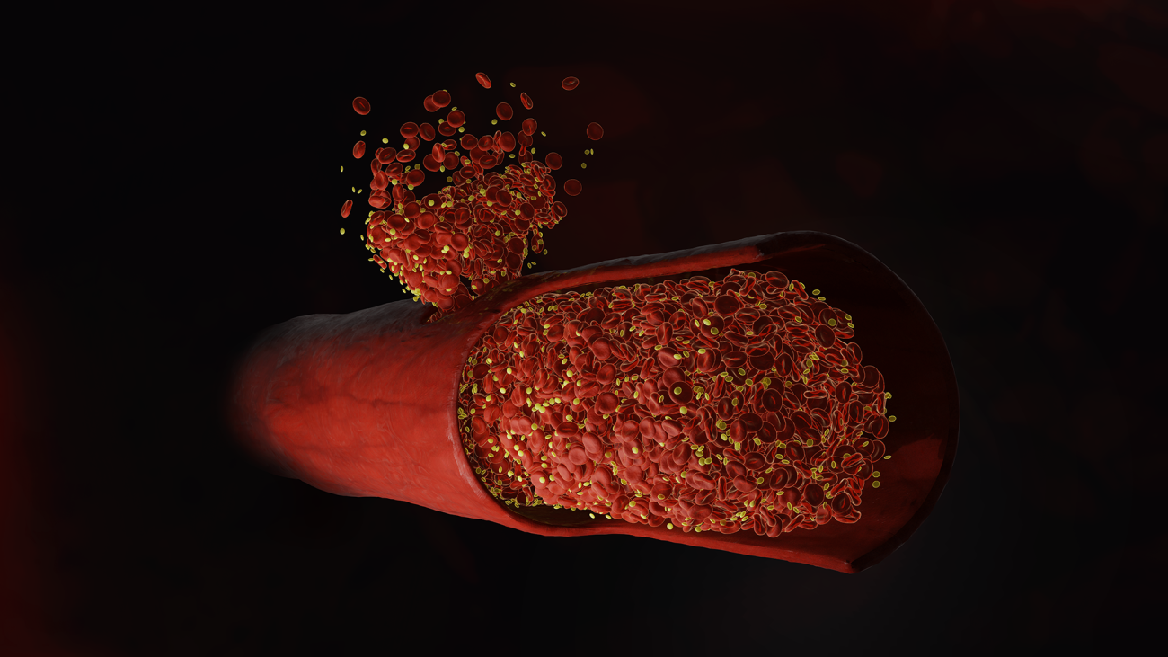
The image depicts a simulation replicating the flow of blood cells in a vessel punctured by a microneedle, similar to a mosquito bite-sized injury. This simulation was conducted to enhance understanding of the blood clotting mechanism.
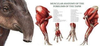
It is based on dissections of tapir limbs and is the first documentation of tapir forelimb anatomy of this accuracy. I strive to compile a whole tapir anatomy atlas since these fascinating and important animals are sparsely studied and poorly understood.

The microscope's laser gazes deep into an Arabidopsis pistil (female flower organ). There, a dye bathes the cell walls in a crimson fire. The ethereal cyan of the SOSEKI5 protein illuminates two chains of ovules (primordial seeds). On the right, hair-like stigma (pollen catcher) cells shimmer in a faint cyan.
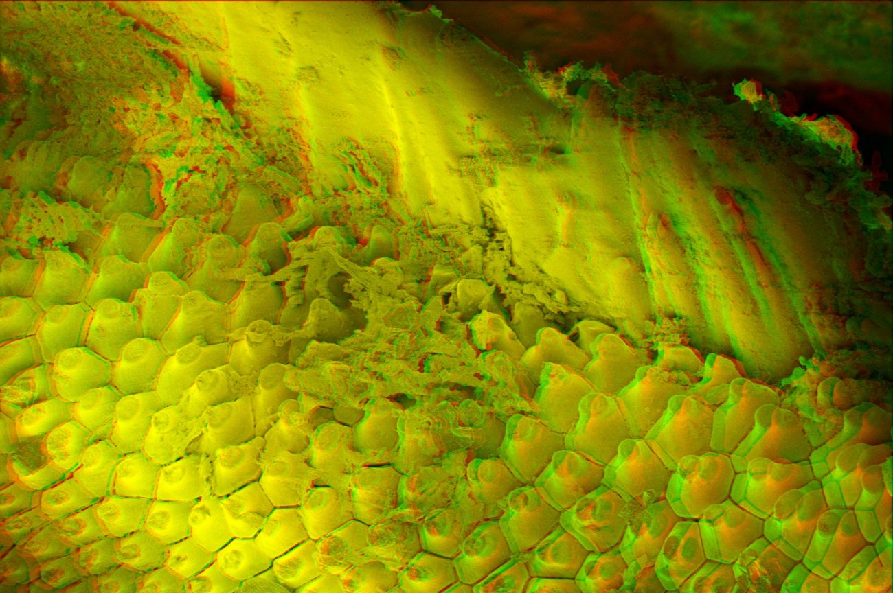
3D Scanning Electron Microscopy image of a cuttlefish suction cup zoomed in on the papillae ring which probably serves to adhere to a relatively rough surface.
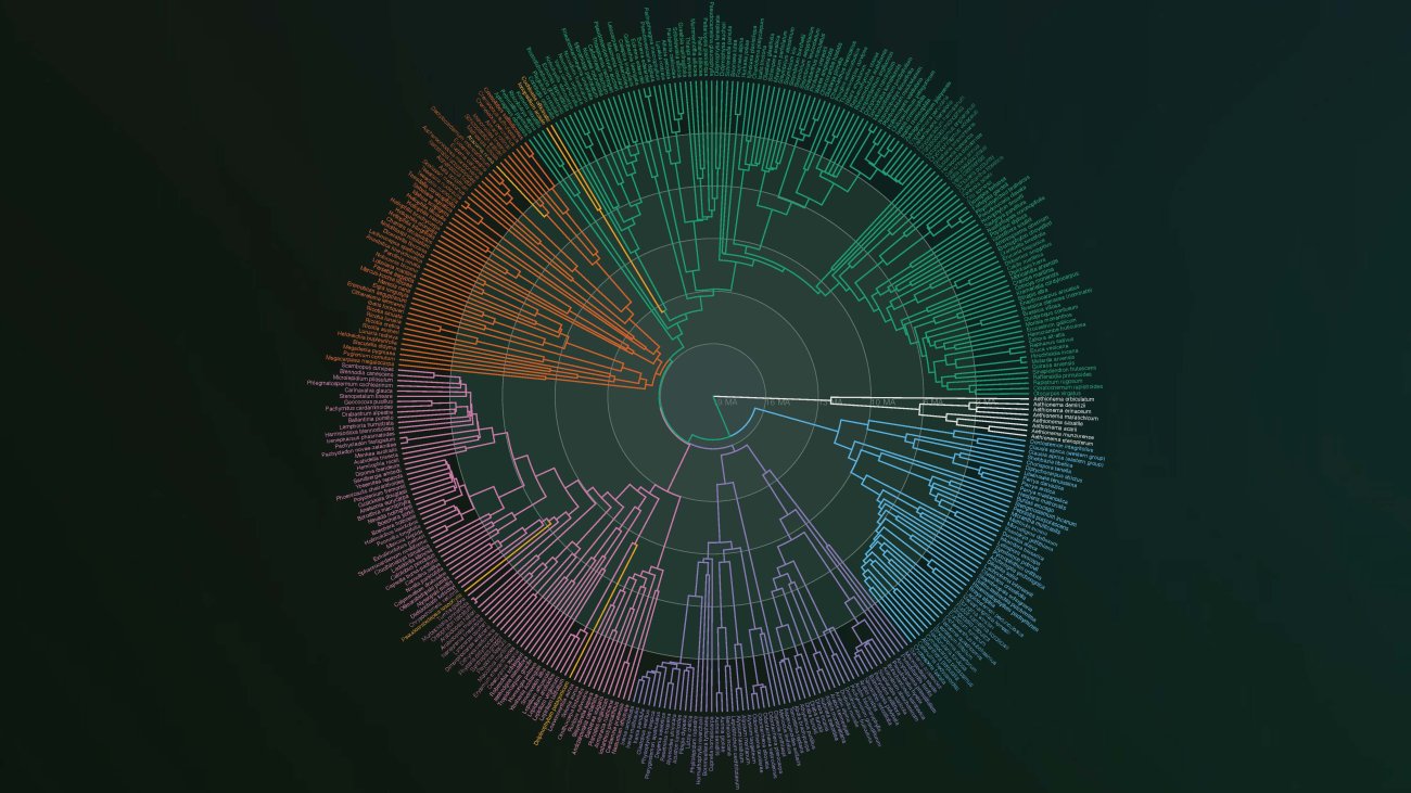
The Tree of Life of the Brassicaceae (BrassiToL), an economically (cabbage, rapeseed) and scientifically (Arabidopsis thaliana) important plant family. The network represents 30 million years of time and family history. The figure is scientifically accurate and can already be explored and questioned online via https://brassitol.vercel.app/.
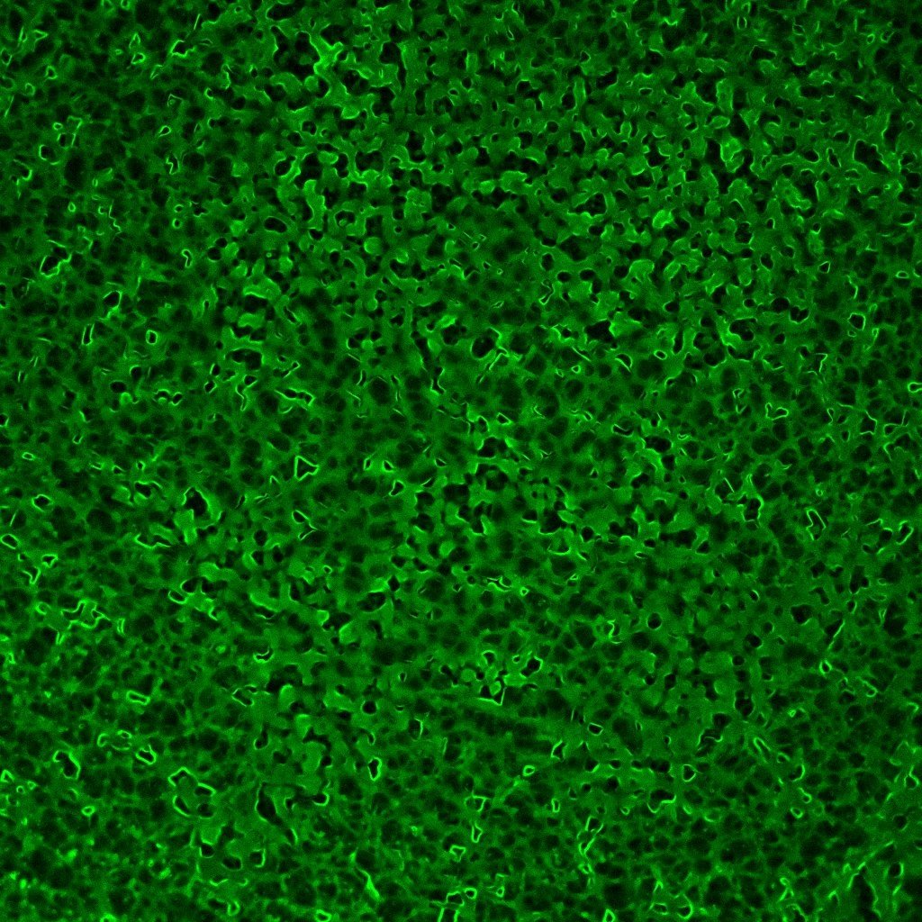
This mesmerising photo shows the phase separation of a tick salivary protein (GRP) manifested into a stunning pattern. A droplet of a buffered GRP solution (containing fluorescently labelled GRP for better visualisation) was allowed to evaporate to induce phase separation, and captured perfectly to reveal protein-rich (bright green) interconnected domains.
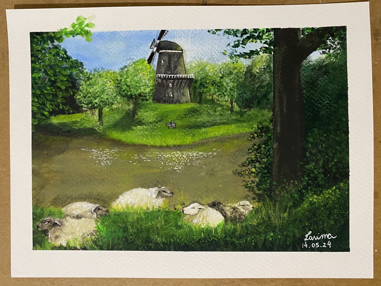
In the picture you see a painting (acrylic on watercolor paper) of a Dutch Landscape. The inspiration of the drawing came from a picture taken in Middelburg, where it is seen the beautiful combination of nature and sheep.